Developmental timeline
TS1 (fertilized egg) → P56 (adult) · click a stage to load samples
TS1
E0.5
1-cell
TS2
E1
2-cell
TS3
E2
4-16 cell
TS4
E3
morula → blastocyst
TS5
E4
late blastocyst
TS6
E4.5
attaching blastocyst
 1
1TS7
E5
egg cylinder
 1
1TS8
E6
prestreak
 2
2TS9
E6.5
streak
 3
3TS10
E7
amnion
 5
5TS11
E7.5
neural plate
 10
10TS12
E8
neural folds
 4
4TS13
E8.5
5-8 somites
 3
3TS14
E9
9-12 somites
 16
16TS15
E9.5
13-20 somites
 6
6TS16
E10
forelimb buds
 8
8TS17
E10.5
hindlimb buds
 7
7TS18
E11
handplate
 15
15TS19
E11.5
hindlimb handplate
 9
9TS20
E12
digital rays
 7
7TS21
E13.5
finger separation
 9
9TS22
E14
eyelid formation
 21
21TS23
E15
eyelids closing
 24
24TS24
E15.5
ear pinna
 5
5TS25
E16.5
long whiskers
 2
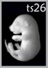
2TS26
E17.5
body hair
 1
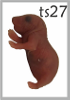
1TS27
E18.5
pre-birth
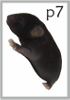
P7
P7
postnatal day 7
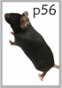
P56
P56
adult (8 weeks)
E0
E2
E4
E6
E8
E10
E12
E14
E16
E18
Birth
E0E5E10E15BirthP7P56
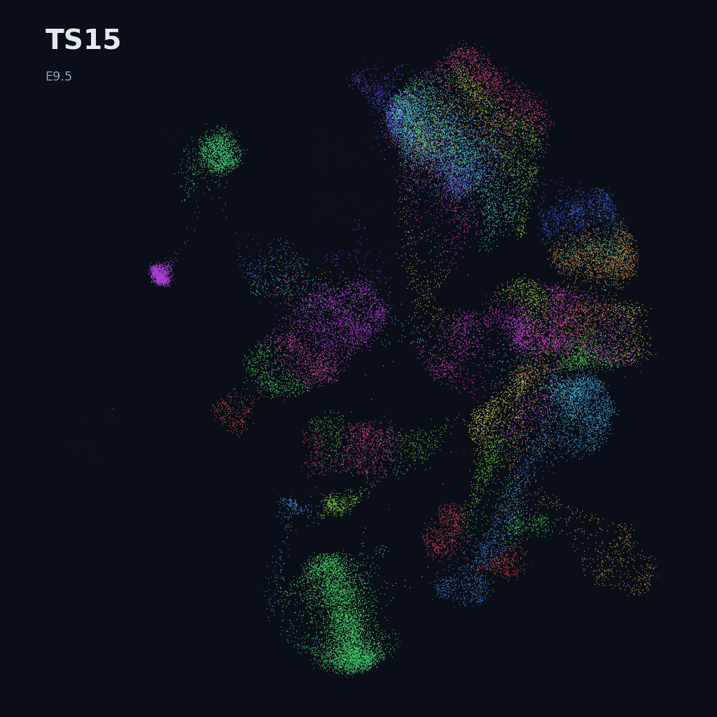
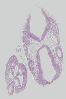
TS15
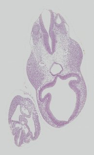
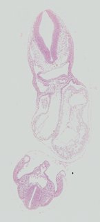
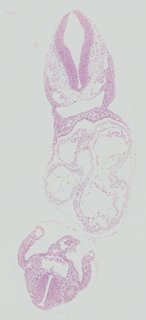
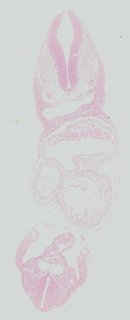
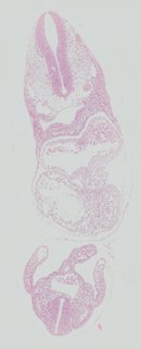
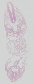
E9.5· 13–20 somitesForelimb field, branchial arches II–III, posterior neuropore still open.
7samples
2/7anatomy delineated
OPT · 7Optical Projection Tomography — a 3D scan of a chemically cleared embryo, captured by rotating it through 360° in a wide-field microscope. Isotropic voxels; raw bg = dark, tissue = bright. Stereo-seq · 7Stereo-seq (BGI) — DNB-array spatial transcriptomics with subcellular ~500 nm resolution. Captures whole-organ to whole-embryo sections; in the Spateo Cell 2024 atlas, 74 sections per embryo were aligned into a 3D cell-bin volume. 10x Chromium v2/v3 + Smart-seq2 · 110x Chromium v2/v3 + Smart-seq2 sci-RNA-seq3 · 1sci-RNA-seq3 — high-throughput single-nucleus combinatorial-indexing RNA-seq (Shendure lab). The OMG mouse prenatal time-lapse profiled 11.4M nuclei across 45 developmental bins from E8 to P0 with this protocol.
Transcriptomics data · 9
Stage-matched · not registered to atlas anatomy Stereo-seq
e9_5_embryo
646,893 cells · 17,649 genes
Spatial · mouse · stage TS15
Qiu et al., Cell 2024 (Spateo)
Open viewer →
Stereo-seq
e9_5_heart
19,780 cells · 12,581 genes
Spatial · mouse · stage E9.5
Qiu et al., Cell 2024 (Spateo)
Open viewer →
Stereo-seq
mosta_e9_5_e1s1
5,913 cells · 25,568 genes
Spatial · mouse · stage TS15
Chen et al., Cell 2022 (MOSTA)
Open viewer →
Stereo-seq
mosta_e9_5_e2s1
5,292 cells · 23,756 genes
Spatial · mouse · stage TS15
Chen et al., Cell 2022 (MOSTA)
Open viewer →
Stereo-seq
mosta_e9_5_e2s2
4,356 cells · 24,107 genes
Spatial · mouse · stage TS15
Chen et al., Cell 2022 (MOSTA)
Open viewer →
Stereo-seq
mosta_e9_5_e2s3
5,059 cells · 24,238 genes
Spatial · mouse · stage TS15
Chen et al., Cell 2022 (MOSTA)
Open viewer →
Stereo-seq
mosta_e9_5_e2s4
5,797 cells · 23,398 genes
Spatial · mouse · stage TS15
Chen et al., Cell 2022 (MOSTA)
Open viewer →
 10x Chromium v2/v3 + Smart-seq2
Extended Mouse Atlas · E6.5–E9.5 gastrulation + early organogenesis
430,339 cells · 27,669 genes (full atlas)
Single-cell · pre-filtered to TS15 · spans 7 stages
Imaz-Rosshandler et al., Development 2024 (Extended Mouse Atlas)
Open UMAP for TS15 →
 sci-RNA-seq3
Mouse prenatal time-lapse · whole atlas
11,441,407 cells · 45,525 genes (full atlas)
Single-cell · pre-filtered to TS15 · spans 16 stages
Qiu et al., Nature 2024 (OMG)
Open UMAP for TS15 →
3D reference models
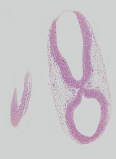
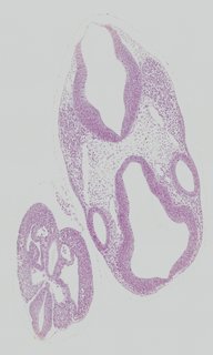
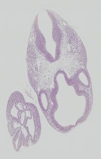
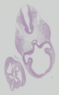
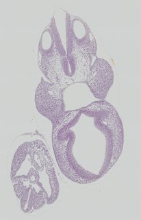
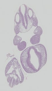
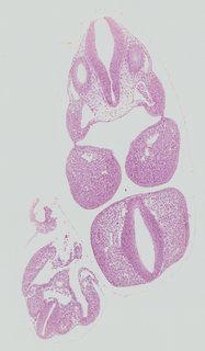
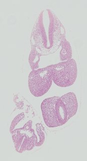
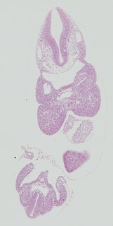
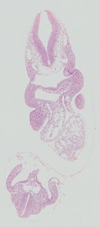
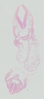
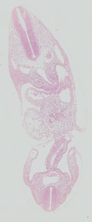
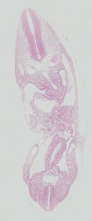
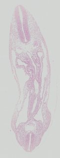
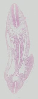
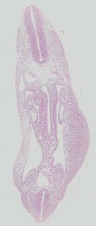
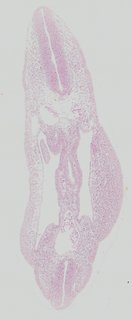
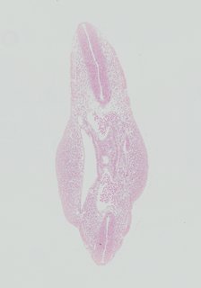
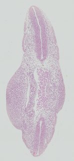
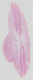
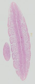
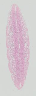
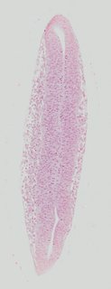
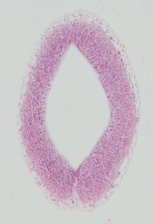
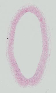
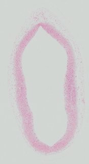
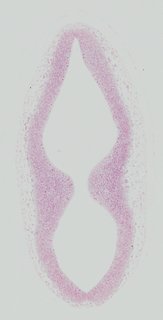
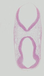
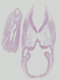
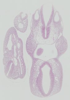
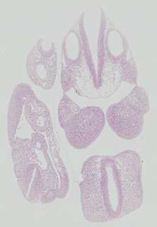
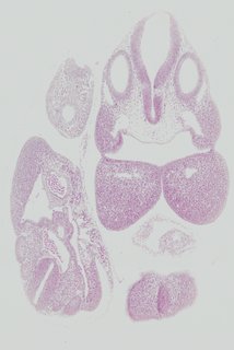
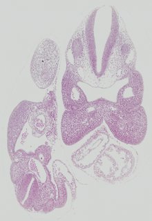
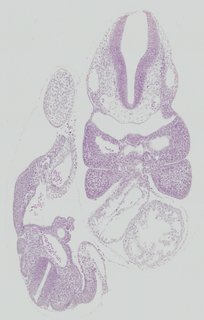
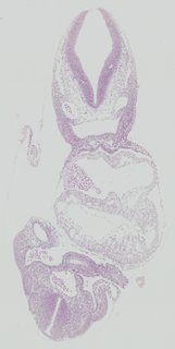
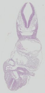
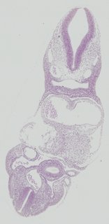
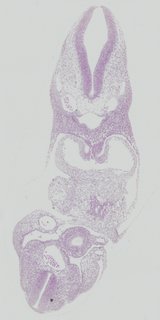
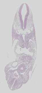
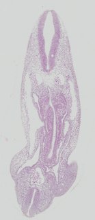
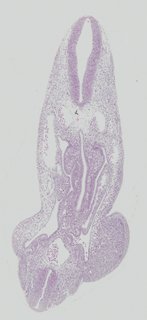
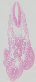
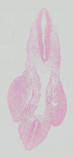
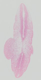
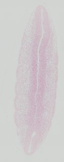
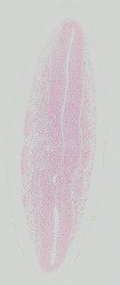
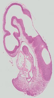
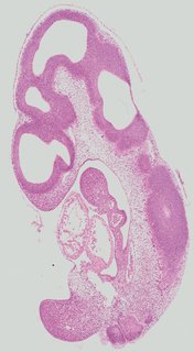
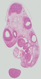
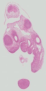
Histology plates · 80
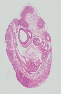
Kaufman atlas, EMAPA-annotated · scroll → 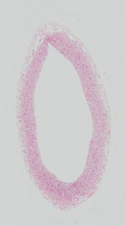 Transverse4 ann
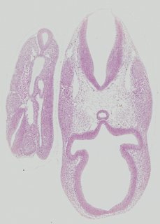
 Plate 19a
TS15
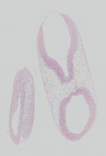
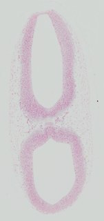
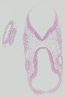
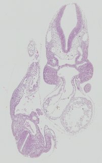
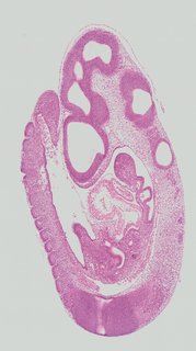
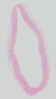 Transverse1 ann
 Plate 19b
TS15
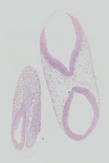
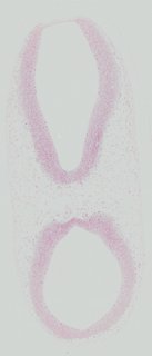
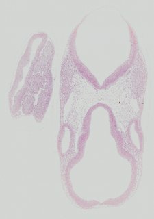
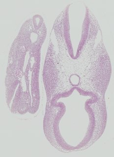
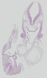
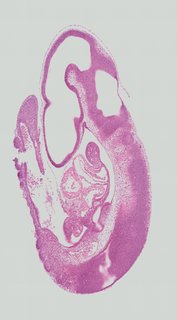
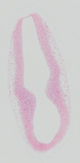 Transverse1 ann
 Plate 19c
TS15
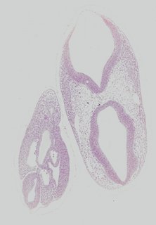
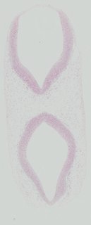
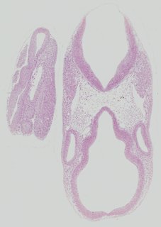
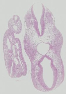
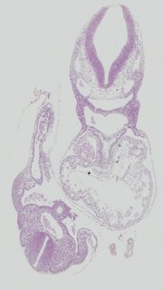
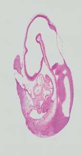
 Transverse5 ann
 Plate 19d
TS15
 Transverse1 ann
 Plate 19e
TS15
 Transverse3 ann
 Plate 19f
TS15
 Transverse10 ann
 Plate 19g
TS15
 Transverse7 ann
 Plate 19h
TS15
 Transverse7 ann
 Plate 19i
TS15
 Transverse3 ann
 Plate 19j
TS15
 Transverse7 ann
 Plate 19k
TS15
 Transverse7 ann
 Plate 19l
TS15
 Transverse17 ann
 Plate 19a
TS15
 Transverse14 ann
 Plate 19b
TS15
 Transverse8 ann
 Plate 19c
TS15
 Transverse6 ann
 Plate 19d
TS15
 Transverse9 ann
 Plate 19e
TS15
 Transverse6 ann
 Plate 19f
TS15
 Transverse8 ann
 Plate 19g
TS15
 Transverse5 ann
 Plate 19h
TS15
 Transverse5 ann
 Plate 19i
TS15
 Transverse5 ann
 Plate 19j
TS15
 Transverse5 ann
 Plate 19k
TS15
 Transverse9 ann
 Plate 19l
TS15
 Transverse24 ann
 Plate 19a
TS15
 Transverse10 ann
 Plate 19b
TS15
 Transverse9 ann
 Plate 19c
TS15
 Transverse4 ann
 Plate 19d
TS15
 Transverse7 ann
 Plate 19e
TS15
 Transverse4 ann
 Plate 19f
TS15
 Transverse4 ann
 Plate 19g
TS15
 Transverse2 ann
 Plate 19h
TS15
 Transverse8 ann
 Plate 19i
TS15
 Transverse7 ann
 Plate 19j
TS15
 Transverse2 ann
 Plate 19k
TS15
 Transverse2 ann
 Plate 19l
TS15
 Transverse4 ann
 Plate 20a
TS15
 Transverse1 ann
 Plate 20b
TS15
 Transverse1 ann
 Plate 20c
TS15
 Transverse1 ann
 Plate 20d
TS15
 Transverse1 ann
 Plate 20e
TS15
 Transverse3 ann
 Plate 20f
TS15
 Transverse2 ann
 Plate 20g
TS15
 Transverse2 ann
 Plate 20h
TS15
 Transverse7 ann
 Plate 20i
TS15
 Transverse6 ann
 Plate 20j
TS15
 Transverse3 ann
 Plate 20k
TS15
 Transverse10 ann
 Plate 20l
TS15
 Transverse20 ann
 Plate 20a
TS15
 Transverse12 ann
 Plate 20b
TS15
 Transverse8 ann
 Plate 20c
TS15
 Transverse9 ann
 Plate 20d
TS15
 Transverse16 ann
 Plate 20e
TS15
 Transverse7 ann
 Plate 20f
TS15
 Transverse13 ann
 Plate 20g
TS15
 Transverse8 ann
 Plate 20h
TS15
 Transverse9 ann
 Plate 20i
TS15
 Transverse6 ann
 Plate 20j
TS15
 Transverse7 ann
 Plate 20k
TS15
 Transverse7 ann
 Plate 20l
TS15
 Transverse25 ann
 Plate 20a
TS15
 Transverse4 ann
 Plate 20b
TS15
 Transverse7 ann
 Plate 20c
TS15
 Transverse14 ann
 Plate 20d
TS15
 Transverse16 ann
 Plate 20e
TS15
 Transverse5 ann
 Plate 20f
TS15
 Transverse4 ann
 Plate 20g
TS15
 Transverse4 ann
 Plate 20h
TS15
 Transverse5 ann
 Plate 20i
TS15
 Transverse6 ann
 Plate 20j
TS15
 Transverse4 ann
 Plate 20k
TS15
 Transverse3 ann
 Plate 20l
TS15
 Sagittal25 ann
 Plate 21a
TS15
 Sagittal19 ann
 Plate 21b
TS15
 Sagittal12 ann
 Plate 21c
TS15
 Sagittal11 ann
 Plate 21d
TS15
 Sagittal24 ann
 Plate 21a
TS15
 Sagittal17 ann
 Plate 21b
TS15
 Sagittal15 ann
 Plate 21c
TS15
 Sagittal11 ann
 Plate 21d
TS15
Knowledge graph · TS15
EMAPA / CL-anchored · drag to rotate, scroll to zoom scRNA-seq · Literature · Predictions — coming soon
Previous
TS14E9
9–12 somites
About Theiler staging
Karl Theiler's staging system (1972) partitions prenatal development into 28 morphologically distinct steps. Virtual Embryo aligns all imagery, anatomy, and future spatial transcriptomics data to this axis.
Next
E10TS16
Forelimb buds
→